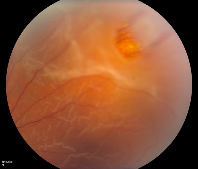
Retinal Detachment
A retinal detachment is separation of the light sensitive retina from the wall of the eye. This results in a loss of vision in the area of separation because the retina is out of focus position and away from the blood supply in the wall of the eye. If the retina is not replaced, the tissue stops working, because of a lack of blood flow, and the vision loss would be permanent. There are 3 major types of retinal detachment. We will discuss the 2 most common forms.
Primary Detachment (Rhegmatogenous)
This is the most common type of retinal detachment (85%). It is caused by a tear or “break” (Rhegmatogenous) in the retina. This tear allows the liquid vitreous to separate the retina from the wall of the eye. A tear is usually caused by the vitreous pulling and tearing the retina. Repair of this type of detachment requires finding the tear(s) and treating them. Treatment may include a gas bubble, laser, scleral buckle or vitrectomy. Ask your doctor which method would fit your situation best.
Tractional Detachment
This type of retinal detachment is caused by pulling on the retina by scar tissue. The most common cause of this type of detachment is diabetes, previous retinal detachment, and scarring. Removal of the scar tissue is the only method to repair the retina since usually there is no hole. Removal of the scar tissue is usually done as part of a Vitrectomy.
Keith Warren, M.D.
Retina Specialist
Board Certified Ophthalmologist
Overland Park and Kansas City